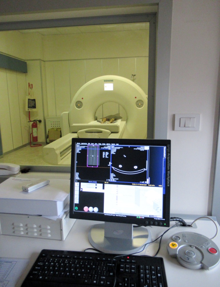
A male individual from the cemetery of Vetricella shows an evident well-healed amputation of the distal end of the right lower limb. Amputation appears to be the result of medical treatment and cases, such as these of survival with an amputated limb in pre-antibiotic era are rare in the anthropological literature. For this study it is essential to analyze the CT (computed tomography) of the lower limbs, recently acquired with a Siemens SOMATOM Scope CT scanner (Fig.1-2).
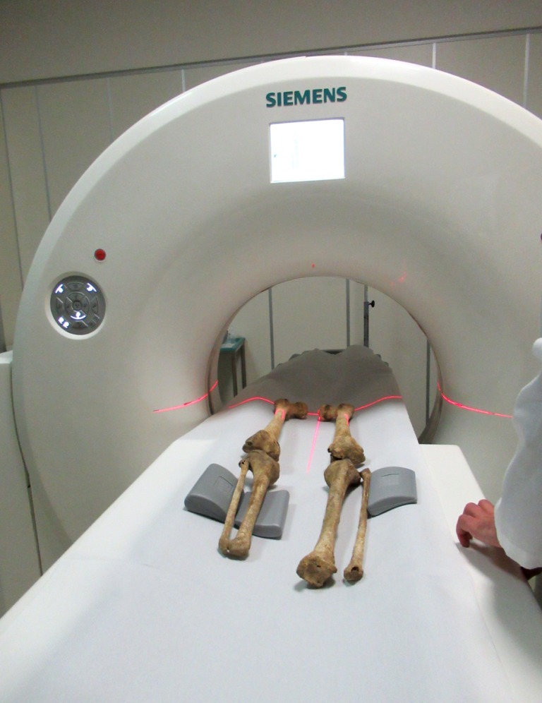
Images of each section of the right and left diaphyses of the femur, tibia and fibula will be measured to compare the diaphyses perimeter, total cross sections areas, cortical thickness and cortical area. Cortical thicknesses will be measured at the four anatomical quadrants (anterior, posterior, medial and lateral). Comparing the data between right and left limb, it will be possible to test the reduced mobility of the amputated limb and eventually hypothesize or exclude the use of prosthesis.